
Bestand:Aphis rosae segmentation.jpg

Oorspronkelijk bestand (935 × 387 pixels, bestandsgrootte: 345 kB, MIME-type: image/jpeg)
| Dit is een bestand van Wikimedia Commons. Onderstaande beschrijving komt van de beschrijving van het bestand daar. |
Beschrijving
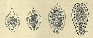
| Beschrijving | FIG. 57. SEGMENTATION OF APHIS ROSAE. (Copied from Metschnikoff.) In all the stages there is seen to be a central yolk mass surrounded by a layer of protoplasm. In this protoplasm two nuclei have appeared in 1, four nuclei in 2. In 3 the nuclei have arranged themselves regularly, and in 4 the protoplasm has become divided into a number of columnar cells corresponding to the nuclei. w. pole of the blastoderm which has no share in forming the embryo |
| Datum | |
| Bron |
https://archive.org/details/theworks02balfuoft/page/116/mode/2up?view=theater&q=blastoderm THE WORKS OF FRANCIS MAITLAND BALFOUR VOL. III A TREATISE ON COMPARATIVE EMBRYOLOGY. |
| Auteur | M. FOSTER, F.R.S., ADAM SEDGWICK, M.A., |
Licentie
|
Dit werk bevindt zich in het publiek domein in landen en gebieden waar de auteursrechttermijn het leven van de auteur plus 70 jaar of minder is.
| |
| Van dit bestand is vastgesteld dat er geen bekende auteursrechtaanspraken op rusten, alle aanverwante en naburige rechten daarbij inbegrepen. | |

|
Dit bestand, dat oorspronkelijk toegevoegd was op een externe website, is nog niet beoordeeld door een moderator of reviewer om te bevestigen dat de opgegeven licentie geldig is. Zie Category:License review needed voor meer instructies.
|
Bestandsgeschiedenis
Klik op een datum/tijd om het bestand te zien zoals het destijds was.
| Datum/tijd | Miniatuur | Afmetingen | Gebruiker | Opmerking | |
|---|---|---|---|---|---|
| huidige versie | 1 mrt 2024 00:34 | | 935 × 387 (345 kB) | Rasbak | {{Information |description=FIG. 57. SEGMENTATION OF APHIS ROSAE. (Copied from Metschnikoff.) In all the stages there is seen to be a central yolk mass surrounded by a layer of protoplasm. In this protoplasm two nuclei have appeared in 1, four nuclei in 2. In 3 the nuclei have arranged themselves regularly, and in 4 the protoplasm has become divided into a number of columnar cells corresponding to the nuclei. w. pole of the blastoderm which has no share in forming the embryo |source=https://ar... |
Bestandsgebruik
Dit bestand wordt op de volgende pagina gebruikt: