
Bestand:Bundleofhis.png
Bundleofhis.png (400 × 483 pixels, bestandsgrootte: 69 kB, MIME-type: image/png)
| Dit is een bestand van Wikimedia Commons. Onderstaande beschrijving komt van de beschrijving van het bestand daar. |
Beschrijving
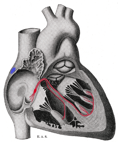
Schematic representation of the atrioventricular bundle of His. The bundle, represented in red, originates near the orifice of the coronary sinus, undergoes slight enlargement to form the AV node. The AV node tapers down into the bundle of HIS, which passes into the ventricular septum and divides into two bundle branches, the left and right bundles. Sometimes the 'left and right bundles of His' are called Purkyne or Purkinge fibres. The ultimate distribution cannot be completely shown in this diagram.
This image is misleading. Although it correctly places the SA and AV nodes in the right atrium, it appears as though there are only two papillary muscles in the right ventricle and three in the left ventricle. The opposite is actually true. The papillary muscles attach to chordae tendinae which then attach to the leaflets of the AV valves, preventing prolapse. The left AV valve is the mitral or bicuspid and only has two leaflets and therefore two papillary muscles. The right AV valve is the tricuspid and should have three papillary muscles corresponding to the three leaflets of the valve.
Licentie
Dit mediabestand is in het publiek domein in de Verenigde Staten van Amerika. Dit geldt voor werken uit de VS waarvan de copyright is verlopen, meestal omdat de eerste publicatie ervan verscheen voor 1 januari 1929. Zie ook deze pagina voor verdere uitleg
|
 | |
 |
Let op! Dit bestand hoeft niet in het publiek domein te zijn buiten de VS (dit geldt voor Canada, China (excl. Hong Kong, Macao of Taiwan), Duitsland, Mexico en Zwitserland). De auteur en het jaar van publicatie zijn essentiële informatie en moeten zijn vastgesteld. Zie ook Wikipedia:Public domain en Wikipedia:Copyrights voor meer informatie.
|
Bestandsgeschiedenis
Klik op een datum/tijd om het bestand te zien zoals het destijds was.
| Datum/tijd | Miniatuur | Afmetingen | Gebruiker | Opmerking | |
|---|---|---|---|---|---|
| huidige versie | 20 sep 2006 19:40 | | 400 × 483 (69 kB) | Kauczuk | Bundle of His, from Gray's Anatomy 1918 |
Bestandsgebruik
Dit bestand wordt op de volgende pagina gebruikt:
Globaal bestandsgebruik
De volgende andere wiki's gebruiken dit bestand:
- Gebruikt op ar.wikipedia.org
- Gebruikt op az.wikipedia.org
- Gebruikt op bn.wikibooks.org
- Gebruikt op bs.wikipedia.org
- Gebruikt op ca.wikipedia.org
- Gebruikt op cs.wikipedia.org
- Gebruikt op de.wikipedia.org
- Gebruikt op de.wikibooks.org
- Gebruikt op el.wikipedia.org
- Gebruikt op en.wikipedia.org
- Gebruikt op en.wikibooks.org
- Gebruikt op es.wikipedia.org
- Gebruikt op es.wikibooks.org
- Gebruikt op eu.wikipedia.org
- Gebruikt op fr.wikipedia.org
- Gebruikt op hi.wikipedia.org
- Gebruikt op it.wikipedia.org
- Gebruikt op ja.wikipedia.org
- Gebruikt op ja.wikibooks.org
- Gebruikt op ko.wikipedia.org
- Gebruikt op lv.wikipedia.org
- Gebruikt op pl.wikipedia.org
- Gebruikt op pt.wikipedia.org
- Gebruikt op sr.wikipedia.org
- Gebruikt op www.wikidata.org