
Bestand:Gray491.png
Gray491.png (500 × 438 pixels, bestandsgrootte: 63 kB, MIME-type: image/png)
| Dit is een bestand van Wikimedia Commons. Onderstaande beschrijving komt van de beschrijving van het bestand daar. |
Beschrijving
| Beschrijving |
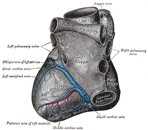
Deutsch: Blick von hinten auf das Herz. Darstellung von Henry Gray. |
||||||||||||||||||||
| Plaat | 491 | ||||||||||||||||||||
| Datum | voor 1858 | ||||||||||||||||||||
| Bron |
|
||||||||||||||||||||
| Auteur |
|
||||||||||||||||||||
.jpg)
Boek
| Henry Gray: Gray's Anatomy (20e editie)
|
|||||||||||||||||||||||
|---|---|---|---|---|---|---|---|---|---|---|---|---|---|---|---|---|---|---|---|---|---|---|---|
| Auteur |
|
-_Title_page.png) | |||||||||||||||||||||
| Bewerker |
Revised by Warren H. Lewis |
||||||||||||||||||||||
| Illustrator |
|
||||||||||||||||||||||
| Titel | |||||||||||||||||||||||
| Uitgave |
20 |
||||||||||||||||||||||
| Uitgever | |||||||||||||||||||||||
| Soort object |
editie, versie of vertaling |
||||||||||||||||||||||
| Pagina's | list of all the plates | ||||||||||||||||||||||
| Taal |
Engels |
||||||||||||||||||||||
| Datum van uitgave |
1918 |
||||||||||||||||||||||
| Plaats van uitgave |
Philadelphia / New York |
||||||||||||||||||||||
| Bron | Bartleby | ||||||||||||||||||||||

Licentie
Deze afbeelding bevindt zich in het publiek domein omdat het een vooral mechanische scan of fotokopie is van een origineel dat zich in het publiek domein bevindt, of – uit het beschikbare bewijs – dat het zo zodanig overeenstemt met deze scan of fotokopie dat er geen copyrightbescherming verwacht kan worden te ontstaan. Het origineel zelf bevindt zich in het publieke domein om de volgende reden:
Deze tag is ontworpen voor gebruik op plaatsen waar er een behoefte is om te verklaren dat alle verbeteringen (bijv. helderheid, contrast, kleur-matching, verscherpen) op zichzelf onvoldoende creatief zijn om een nieuw auteursrecht te genereren. Het kan gebruikt worden wanneer het niet bekend is of er verbeteringen zijn aangebracht, en wanneer de verbeteringen duidelijk maar onvoldoende zijn. Voor bekende ruwe onverbeterde scans kunt u gebruik maken van een geschikte {{PD-old}}-tag in plaats daarvan. Voor gebruik, zie Commons:When to use the PD-scan tag.  | ||||
The coronary sinus is a collection of veins joined together to form a large vessel that collects blood from the myocardium of the heart. It is present in humans and other animals. It delivers deoxygenated blood to the Right atrium in conjunction with the superior and inferior vena cava.
The coronary sinus opens into the right atrium, between the inferior vena cava and the atrio-ventricular orifice. It returns the blood from the substance of the heart, and is protected by a semicircular fold of the lining membrane of the auricle, the coronary valve (the valve of Thebesius). The sinus, before entering the auricle, is considerably dilated - nearly to the size of the end of the little finger. Its wall is partly muscular, and at its junction with the great coronary vein is somewhat constricted and furnished with a valve consisting of two unequal segments.(Gray 462)
Location: It is located in the right atrium and runs transversely in the groove between the left atrium and ventricle on the posterior surface of the heart.
The coronary sinus orifice (opening) is just superior to the septal leaflet of the tricuspid valve. The coronary sinus orifice is also known as the ostium of the coronary sinus, and is guarded by the Thebesian valve.
Drainage: It receives blood mainly from the small, middle, great and oblique cardiac veins. It also receives blood from the left marginal vein and the left posterior ventricular vein. The anterior cardiac veins drain directly into the right atrium. (Some small veins drain into any of the four chambers of the heart.)
It drains into the right atrium on the posterior, inferior surface, medial to the inferior vena cava opening.
Bestandsgeschiedenis
Klik op een datum/tijd om het bestand te zien zoals het destijds was.
| Datum/tijd | Miniatuur | Afmetingen | Gebruiker | Opmerking | |
|---|---|---|---|---|---|
| huidige versie | 23 jan 2007 22:35 | | 500 × 438 (63 kB) | Pngbot | optimized with optipng |
| 11 feb 2006 08:26 |  | 500 × 438 (100 kB) | Arcadian | {{Gray's Anatomy plate}} |
Bestandsgebruik
Dit bestand wordt op de volgende 2 pagina's gebruikt:
Globaal bestandsgebruik
De volgende andere wiki's gebruiken dit bestand:
- Gebruikt op ar.wikipedia.org
- Gebruikt op bg.wikipedia.org
- Gebruikt op bn.wikipedia.org
- Gebruikt op bs.wikipedia.org
- Gebruikt op cv.wikipedia.org
- Gebruikt op de.wikibooks.org
- Gebruikt op el.wikipedia.org
- Gebruikt op en.wikipedia.org
- Coronary circulation
- Coronary sinus
- Oblique vein of the left atrium
- Posterior descending artery
- Circumflex branch of left coronary artery
- Vital heat
- Posterior interventricular sulcus
- Left marginal artery
- Smallest cardiac veins
- Vascular remodelling in the embryo
- Crux cordis
- User:Bob K31416/BH
- User:Walkerc84/sandbox
- User:Was a bee/Gray
- Gebruikt op es.wikipedia.org
- Gebruikt op fa.wikipedia.org
- Gebruikt op it.wikipedia.org
- Gebruikt op ja.wikipedia.org
- Gebruikt op ko.wikipedia.org
- Gebruikt op nn.wikipedia.org
- Gebruikt op pl.wikipedia.org
- Gebruikt op pt.wikipedia.org
- Gebruikt op ro.wikipedia.org
Globaal gebruik van dit bestand bekijken.