
Bestand:Nicotiana tabacum fpls-12-642879-g002.jpg

Grootte van deze voorvertoning: 497 × 599 pixels. Andere resoluties: 199 × 240 pixels | 398 × 480 pixels | 637 × 768 pixels | 849 × 1.024 pixels | 2.126 × 2.564 pixels.
Oorspronkelijk bestand (2.126 × 2.564 pixels, bestandsgrootte: 1,06 MB, MIME-type: image/jpeg)
| Dit is een bestand van Wikimedia Commons. Onderstaande beschrijving komt van de beschrijving van het bestand daar. |
Beschrijving
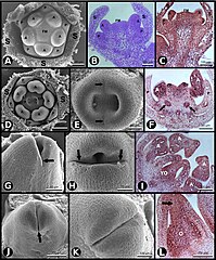
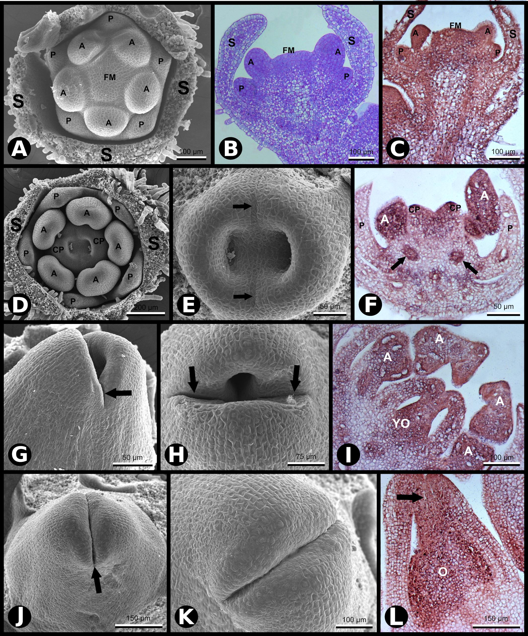
| Beschrijving | Figure 2. SCI1 expression during later stages of floral development (continuation of the stages shown in Figure 1). (A) SEM of a flower bud in which petals and anthers are emerging (advanced stage –8). Scale bar: 100 μm. (B) Bright-field microscopy showing a longitudinal section of a flower bud in a developmental stage equivalent to the one shown in (A). Scale bar: 100 μm. (C) In situ hybridization of a flower bud (advanced stage –8) with SCI1 antisense probe. Scale bar: 100 μm. (D) SEM of a flower bud at stage –7 (as defined by Koltunow et al., 1990), in which carpels are emerging. Scale bar: 200 μm. (E) A higher magnification view of the flower bud shown in (D), in which the fusion lines are visible (arrows). Scale bar: 50 μm. (F) In situ hybridization of a flower bud at stage –7/–6, with SCI1 antisense probe. Arrows point to ovary locules. Scale bar: 50 μm. (G,H) SEM of flower buds at stage –6; carpels fused at the base and not yet fused at the top. Scale bars: 50 μm (G) and 75 μm (H). (I) In situ hybridization of a flower bud at late stage –6, with SCI1 antisense probe. Scale bar: 100 μm. (J,K) SEM of flower buds at stage –5; carpels already fused at the top; the fusion region is a site of intense cell proliferation. Scale bars: 150 μm (J) and 100 μm (K). (L) In situ hybridization with SCI1 antisense probe of a flower bud at late stage –5; style beginning to form (arrow). Scale bar: 150 μm. Floral meristem (FM), sepals (S), petals (P), anther (A), carpels (C), carpel primordia (CP), ovary (O), young ovary (YO). |
| Datum | |
| Bron | https://www.frontiersin.org/files/Articles/642879/fpls-12-642879-HTML/image_m/fpls-12-642879-g002.jpg, SCI1 Is a Direct Target of AGAMOUS and WUSCHEL and Is Specifically Expressed in the Floral Meristematic Cells, Front. Plant Sci., 18 March 2021,Sec. Plant Development and EvoDevo, Volume 12 - 2021, https://doi.org/10.3389/fpls.2021.642879 |
| Auteur | Joelma O. Cruz Juca A. B. San Martin, Greice Lubini, Edward J. Strini1, Rómulo Sobral, Vitor F. Pinoti, Pedro B. Ferreira, Vanessa Thomé, Andréa C. Quiapim, Marcelo C. Dornelas, Maria Cristina, S. Pranchevicius, Francisco Madueño, M. Manuela R. Costa, Maria Helena S. Goldman, |
Open access
Licentie
Dit bestand is gelicenseerd onder de Creative Commons Naamsvermelding 4.0 Internationaal licentie.
- De gebruiker mag:
- Delen – het werk kopiëren, verspreiden en doorgeven
- Remixen – afgeleide werken maken
- Onder de volgende voorwaarden:
- naamsvermelding – U moet op een gepaste manier aan naamsvermelding doen, een link naar de licentie geven, en aangeven of er wijzigingen in het werk zijn aangebracht. U mag dit op elke redelijke manier doen, maar niet zodanig dat de indruk wordt gewekt dat de licentiegever instemt met uw werk of uw gebruik van zijn werk.
Bestandsgeschiedenis
Klik op een datum/tijd om het bestand te zien zoals het destijds was.
| Datum/tijd | Miniatuur | Afmetingen | Gebruiker | Opmerking | |
|---|---|---|---|---|---|
| huidige versie | 30 dec 2023 00:28 | | 2.126 × 2.564 (1,06 MB) | Rasbak | {{Information |description=Figure 2. SCI1 expression during later stages of floral development (continuation of the stages shown in Figure 1). (A) SEM of a flower bud in which petals and anthers are emerging (advanced stage –8). Scale bar: 100 μm. (B) Bright-field microscopy showing a longitudinal section of a flower bud in a developmental stage equivalent to the one shown in (A). Scale bar: 100 μm. (C) In situ hybridization of a flower bud (advanced stage –8) with SCI1 antisense probe. Scale... |
Bestandsgebruik
Dit bestand wordt op de volgende pagina gebruikt: