
Bestand:Spindle centriole - embryonic brain mouse - TEM.jpg

Oorspronkelijk bestand (1.283 × 1.600 pixels, bestandsgrootte: 901 kB, MIME-type: image/jpeg)
| Dit is een bestand van Wikimedia Commons. Onderstaande beschrijving komt van de beschrijving van het bestand daar. |
Beschrijving
| Beschrijving |
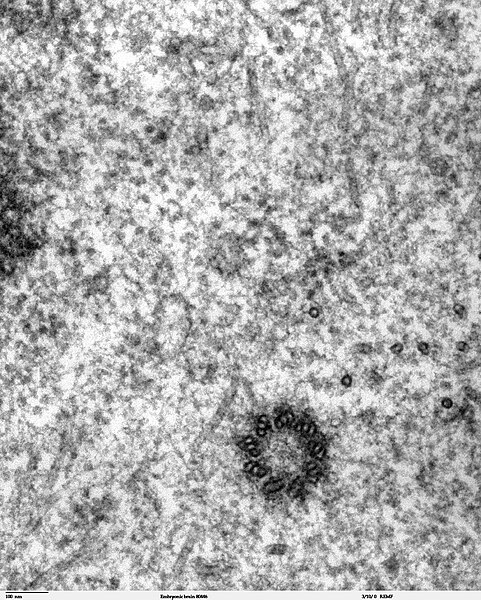
Transmission electron microscope image of a thin section cut through the developing brain tissue (telencephalic hemisphere) of an 11.5 day mouse embryo. This high magnification image of "Embryonic brain 80445" show a spindle centriole and some spindle microtubules visible in the cytoplasm of a mitotic cell at the luminal surface of the telencephalon. JEOL 100CX TEM References: Marin-Padilla, M. (1985) "Early Vascularization of the Embryonic Cerebral Cortex: Golgi and Electron Microscope Studies", J. Comparative Neurology, 241:237-249 Marin-Padilla, M. and M. Amievo (1989) "Early Neurogenesis of the Mouse Olfactory Nerve: Golgi and Electron Microscope Studies", J. Comparative Neurology, 288:339-352 |
| Bron | |
| Auteur | Louisa Howard, Miguel Marin-Padilla |
| Toestemming (Hergebruik van dit bestand) |
PD |
Licentie
| Dit werk vrijgegeven in het publieke domein door de auteur, Louisa Howard, Miguel Marin-Padilla. Dit is wereldwijd van toepassing. In sommige landen is dit wettelijk niet mogelijk; in die gevallen geldt: Louisa Howard, Miguel Marin-Padilla staat iedereen toe dit werk voor eender welk doel te gebruiken, zonder enige voorwaarden, tenzij zulke voorwaarden door de wet worden voorgeschreven.
|
Bestandsgeschiedenis
Klik op een datum/tijd om het bestand te zien zoals het destijds was.
| Datum/tijd | Miniatuur | Afmetingen | Gebruiker | Opmerking | |
|---|---|---|---|---|---|
| huidige versie | 3 nov 2006 00:02 | | 1.283 × 1.600 (901 kB) | Patho | {{Information |Description=Transmission electron microscope image of a thin section cut through the developing brain tissue (telencephalic hemisphere) of an 11.5 day mouse embryo. This high magnification image of "Embryonic brain 80445" show a spindle cen |
Bestandsgebruik
Dit bestand wordt op de volgende pagina gebruikt:
Globaal bestandsgebruik
De volgende andere wiki's gebruiken dit bestand:
- Gebruikt op ar.wikipedia.org
- Gebruikt op bg.wikipedia.org
- Gebruikt op bs.wikipedia.org
- Gebruikt op ca.wikipedia.org
- Gebruikt op cs.wikipedia.org
- Gebruikt op da.wikipedia.org
- Gebruikt op de.wikibooks.org
- Gebruikt op en.wikipedia.org
- Gebruikt op en.wikibooks.org
- Gebruikt op es.wikipedia.org
- Gebruikt op eu.wikipedia.org
- Gebruikt op fr.wikipedia.org
- Gebruikt op gl.wikipedia.org
- Gebruikt op gv.wikipedia.org
- Gebruikt op hu.wikipedia.org
- Gebruikt op kk.wikipedia.org
- Gebruikt op nl.wikibooks.org
- Gebruikt op pt.wikipedia.org
- Gebruikt op sv.wikipedia.org
- Gebruikt op th.wikipedia.org
- Gebruikt op tr.wikipedia.org